Ms. Daassi Dhouha, 5th year student, published a first author paper “Differential expression patterns of MafB and c-Maf in macrophages in vivo and in vitro” in Biochemical and Biophysical Research Communications.
Summary for HBP website:
Miss Dhouha Daassi, 5th grade in the Human Biology Program of Ph.D. has published her first first-author paper in in the journal of :Biochemical and Biophysical Research Communications on March 16th 2016.
To generate the organisms, a large network of transcription factors is needed. The distinguished team of miss Dhouha Daassi works on the large Maf proteins group: MafA, MafB, c-Maf and NRL whose main function is to regulate the terminal differentiation in many tissues like bone, brain, kidney, lens, pancreas and retina in addition to blood. The large Maf transcription factors MafB and c-Maf have been previousely reported to have overlapping functions in macrophages. But in some recent reports the opposite can also be inferred. Due to this paradox, miss Daassi aimed to study the expression patterns of MafB and c-Maf in various tissue-resident macrophage types to help understanding their functions.
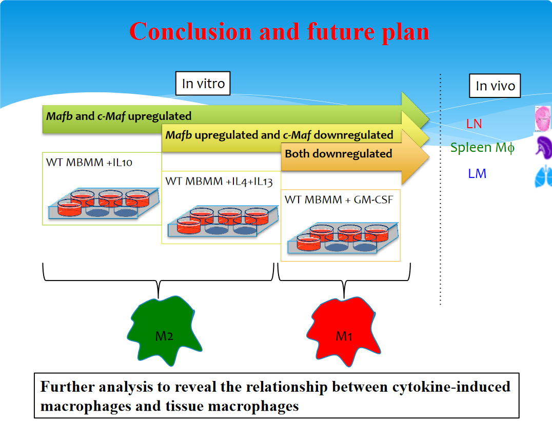
Miss Daassi detected both MafB and c-Maf signals in lymph node macrophages. In vitro part showed that in the splenic macrophages the MafB signal was detected, but the c-Maf signal was not. No expression of c-Maf or MafB was detected in the lung or kidney macrophages. She could confirm by Flow cytometry analysis a similar pattern of GFP expression in Mafb/GFP knock-in heterozygous mice. To analyze these different expression patterns in greater detail, she examined the expression of MafB and c-Maf by quantitative RT-PCR in different cytokine- or LPS-induced macrophages in vitro (M1 and M2 subtypes). Miss Dhouha found that MafB expression was induced by IL-10 or IL-4 with IL-13 and was reduced by LPS or GM-CSF. By contrast, c-Maf expression was induced by IL-10 and reduced by IL-4 with IL-13 or GM-CSF) which means that the two transcription factors are mainly expressed by M2 not M1. Taking together the in vivo and in vitro results, MafB and c-Maf have different expression patterns in tissue-resident macrophages, suggesting differences in function. The following cartoon shows how the in vivo with the vitro study contributed together to shape the whole puzzle of the expression patterns of these two transcription factors. For more details please visit the link below:
http://www.sciencedirect.com/science/article/pii/S0006291X16303758


